Version française
Biobank Data Base
Rapid Tissue Freezing
Imaging
Introduction to Tissue Arrays
Ready-to-use Tissue Arrays
Design of TMAs
Tissue Arrayer MiniCore®
Analysis of TMAs
TISALYS® Data Base
Publications & Links
 
Tissue Array
  Ready-to-use Tissue Arrays


  PETAGEN Slides
AccuMax Array is a slide containing various types of small tissues. The diameter of each spot on the slide ranges from 0.6 to 2.0 mm and the sections number tens to several hundreds
Each tissue section is extracted from various donor blocks and transferred into a ready-made recipient block. The donor blocks are generally conventional, formalin-fixed, paraffin-embedded blocks but can also be different types, such as non-formalin-fixed, frozen types.
Benefits

- Providing every feature of the whole tissue sections
- Allowing the observation of proteins
- RNA and DNA from almost the same region saving you
- Time: several days to even weeks or months
- Money: several hundreds to tens of thousands of dollars
- Labor: one experiment or several hundreds


Minimizing experimental errors caused by different conditions
Who needs AccuMax™ Array ?
Those hoping to :
- Discover various molecular events or molecules ,
- Find candidates for novel molecular targets such as a cancer gene ,
- Validate candidates for novel molecular targets using human tissues with appropriate information .
Why AccuMax™ Arrays are the best slides for the Tissue Array ?
Thorough quality control :
- Almost no loss of tissue spots
- Maximum preservation of tissue antigenicity
- Allows for detailed pathological and clinical information
- Variety of organs that can fit on a single slide
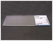
Which are the caratheristics of AccuMax™ Arrays ?
- Formalin-/Paraformaldehyde-fixed or frozen tissues
- Ideal diameter for tissue section spot (1.0 mm)
- Easy-to-read format
- Individually packed to maintain maximum antigenicity and prevent oxidation
- Relevant information and certificates
Quality Assurance & Quality Control
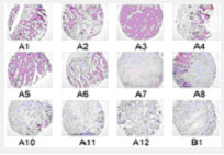
To guarantee the correctness of each tissue spot on the AccuMax Tissue Microarray:
1. Every 20 slides and the last slides from a block are H&E stained and read by a certificated pathologist

2. Randomly selected H&E stained are re- read by another pathologist

3. All Tissue Microarrays are iindividually examined by well trained technologists before packing to guarantee no loss of tissue spots

4.Packages are double checked to find any defects
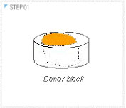
Step 1 : Preparation of a donor block

1. Conducted by certificated pathologists and technologists
2. Through standardized and strict protocol supervised by certificated pathologists
3. Use of top-quality materials
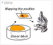
Step 2: Selection of the to-be punched position in the donor block

Conducted and double checked by certificated pathologists
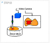
Step 3: Punching the tissue target zone

1. Guided magnified image in real time
2. Carried out to minimize errors from manual operation
3. Specially designed needle to guarantee the smoothness of margin of punched core.
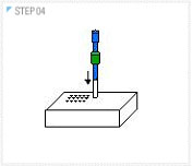
Step 4: Making a hole into which the punched core is going to be transferred

1. Specially designed needle to guarantee the fitness of core to the hole
2. Carried out to minimize errors from manual operation
3. Use fully qualified materials having hardness and minimum crack during process.
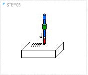
Step 5: Transfer of the core into the hole transfer of the core into the hole

Carried out to minimize errors stemming from manual operation - automatic control of the positionn
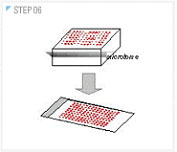
Step 6: Manufacturing the Tissue MicroArray

1. Conducted by certificated and skillful technologists
2. VRegular check-up with instruments and use of top-quality materials
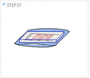
Step 7: Packing the Tissue MicroArray

1. Double-packed individually with two different packing materials under nitrogen atmosphere to prevent oxidation and drying
2. Specially designed packing materials: a hard plastic box and an opaque bag
Step 8: Tracking

All labels are pre-attached to the materials before use and all task carried by two different persons simultaneously
FAQ
01. How are the slides processed, and is there a quality control program for Tissue Array preparation ?
02. What's so special about AccuMax™ Array ?
03. What are the limitations of Tissue Array ?
04. Which information is provided for each tissue specimen ?
01.How are the slides processed, and is there a quality control program for Tissue Array preparation ?

Tissues are processed from the point of fixation in paraformaldehyde until clearing in xylene by a standardized autoprocessor program. Highly-qualified paraffin-embedded blocks are sectioned off by a disposable blade in a microtome. The pathologist differentiates between cancer and pathologic regions in H&E stained slides. Precise input of the exact coordinates into a computer program allows a newly made array block to be constituted by delicately arranging tissue cores. The final QC step is completed by the same pathologists who initially prepared the TMA.
02.What's so special about AccuMax™ Array ?

We have analyzed and addressed nearly every issue related to current TMA slide needs: large numbers of tissues of various types; pathologic tissue alongside of its cancer matching pair from the the same organ; stubborn adherence of tissues to withstand the strain of the staining procedure; regular and precise arrangement of section spots; detailed pathological attached not only to the diseased but also the cancer tissues. And all this at a very reasonable price!
03.What are the limitations of Tissue Array ?

A small tissue core may result in an insufficient number of cells and tissue loss can often occur. To overcome these limitations, as many tissue cores of appropriate size are inserted in a duplicate/triplicate manner as possible under optimal conditions.
04.Which information is provided for each tissue specimen ?
The patient's personal information is kept strictly confidential, but age, sex and diagnosis will be included as a matter of course. We provide a detailed clinical history of each tissue section so that each slide can also be used for scientific research related to the clinical area.
I wish a quotation
I wish more information
top of page